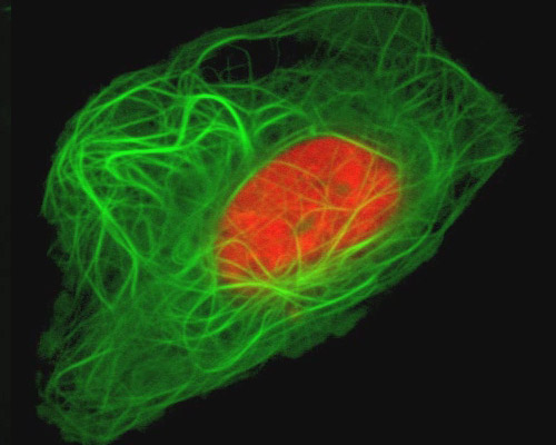
Human Osteosarcoma with mEmerald-MAP4 and mCherry-H2B

Cell nuclei were visualized in human osteosarcoma cells (U-2 line) in the featured digital video sequences with the red fluorescent protein mCherry fused to H2B, a histone protein. Histones, which are closely associated with DNA, are involved in nucleosome formation and in the regulation of chromatin and genes. First discovered by Albrecht Kossel in the late 19th century, histones were not well understood for more than a hundred years. As late as the early 1990s, histones were still commonly thought to be essentially a sort of nuclear packing material.
In addition, microtubules were fluorescently tagged with mEmerald fused to MAP4, a microtubule-associated protein. mEmerald is a green fluorescent variant of EGFP with improved brightness and photostability. Excitation of mEmerald peaks at 487 nanometers and emission peaks at 509 nanometers. The red fluorescent protein employed to tag histone H2B, mCherry, was developed through directed mutagenesis of mRFP1, a monomeric DsRed mutant. Peak excitation and emission of mCherry occur at 587 and 610 nanometers, respectively.


