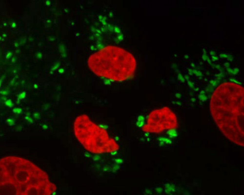
Pig Kidney Cells with mEmerald-Golgi and mCherry-H2B

The Golgi apparatus was one of the first organelles to be identified with a light microscope, as its comparatively large size and regular configuration make it a prominent cellular feature. The Golgi body appears as a group of flattened, sac-like structures called cisternae, each of which is enclosed by a membrane. The number of cisternae in a Golgi stack differs and several stacks may be found in the same cell. In some types of cells, multiple Golgi stacks are connected by tubular linkages. This results in a sizable complex normally located near the centrosome adjacent to the nucleus. The primary function of vesicles concentrated in the area surrounding the Golgi apparatus is to transfer material from the organelle to other cellular structures.
The pig kidney epithelial cells (LLC-PK1 line) featured in this digital video were fluorescently tagged with mCherry fused to the H2B histone protein and mEmerald fused to the Golgi apparatus. mCherry is a red member of the mFruit series of fluorescent proteins derived from mRFP1. Peak excitation and emission wavelengths of mCherry occur at 587 and 610 nanometers, respectively. The green fluorescent protein mEmerald is a bright, photostable variant of enhanced Aequorea GFP. Excitation of mEmerald peaks at 487 nanometers and emission peaks at 509 nanometers.


