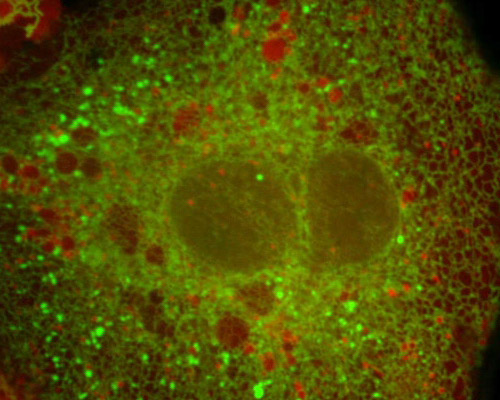
Pig Kidney Cells with mEmerald-ER and mCherry-Rab7a

The red fluorescent protein mCherry has been successfully fused to several proteins, including actin, tubulin, and fascin. Other fusion proteins containing mCherry have been reported in zebrafish, E. coli, HIV virions, and yeast. mCherry is a useful tool because the ample separation in color between mCherry and a green fluorescent protein such as mEmerald provides exceptional conditions for identifying protein interactions inside cells.
Endosomes are membrane-bound vesicles that function in material storage and sorting within cells. Proteins, lipids, and other materials are delivered to the Golgi apparatus, lysosomes, and even the cell surface via endosomes. A number of different complex pathways may be taken by individual endosomes depending on their contents and cellular needs. Late endosomes were tagged in the digital video sequence in this section with mCherry fused to Rab7a.
mCherry was one of the original monomeric “mFruit” fluorescent proteins exhibiting emission maxima ranging from 540 nm to 610 nm developed by a team led by Nathan C. Shaner. The team produced the group of proteins through the directed evolution of mRFP1. mCherry is generally regarded as one of the most useful of the mFruit fluorescent proteins. In addition to endosomes, the featured video examines the endoplasmic reticulum, which was visualized with the EGFP variant mEmerald.


