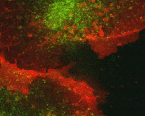
Monkey Kidney Cells with mEGFP-TFR and mCherry-CAAX

The enhanced green fluorescent protein (EGFP) is a transformation marker. EGFP is used for brighter fluorescence and higher expression in mammalian cells. There are more than 190 silent base changes in the coding sequence of the EGFP gene, which correspond to human codon-usage preference. EGFP is expressed in the SV40 enhancer/promoter, whose origin also allows for replication in mammalian cells expressing the SV40 T antigen, such as in the African green monkey kidney epithelial cells (BS-C-1 line). A transferrin receptor was visualized in the digital video sequence in this section with mEGFP, an enhanced version of Aequorea-based GFP. Lysosomes are visualized in the BS-C-1 cells featured in the digital video sequence in this section with mApple.
Transferrin is a trace plasma protein that reversibly binds iron, transports it through the blood, and delivers it to cells. To gain entry into a cell, transferrin must encounter transferrin receptor, the levels of which are regulated by intracellular iron concentration. The transferrin binds to transferrin receptor and is then imported into the cell in a vacuole which is broken down in order to release the iron it contains. The transferrin receptor then follows the endocytic pathway back to the cellís surface in order to import additional iron if needed.


