


Widefield Imaging Applications
Showcase your most challenging applications in widefield fluorescence microscopy together with ZEISS. Investigators are encouraged to upload their images and experimental details, which will be reviewed and integrated into a separate page and displayed on the ZEISS Online Campus. The links provided below highlight widefield fluorescence microscopy applications presented by scientists from around the world.
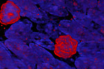
Department of Neurobiology / Yale University
Embryonic SVZ cell (neuronal progenitor) nuclei stained with Ki67 (red) and DAPI (blue).
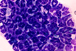
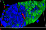
Department of Microbiology, Montana State University
This image shows an aggregate of neutrophils formed in vivo in the mouse lung following exposure to A. fumigatus conidia.
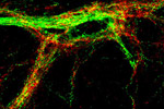
Tufts-New England Medical Center
Double labeling immunofluorescence showing association of TRH-containing axons (red) and cocaine-and amphetamine related transcript (CART)-containing axons (green) in the spinal cord of an adult, male, Sprague-Dawley rat.
Carnegie Institution, Departement of Embryology
A 3D reconstruction of the tip of a drosophila ovary. Nuclei are in blue, follicle cells are in green and one follicle cell (red) is in mitosis.

National Institute of Child Health and Human Development
An 8-cell intestinal stem cell clone (marked in green) made up of a single stem cell (asterisk), recent stem cell daughter known as an enteroblast (adjacent to the stem cell), two enteroendocrine cells (arrowheads), and one early enterocyte (next to the enteroblast) and 3 mature enterocytes (larger nuclei).