


Sclerenchyma Tissue - 40x (Autofluorescence)
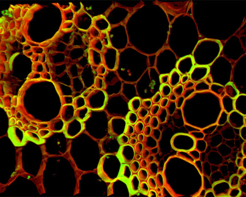
The digital image shown above is a reconstruction of thick tissue sections of sclerenchyma imaged by means of autofluorescence. Sclerenchyma is plant tissue composed of cells with thickened, lignified secondary cell walls that have reached maturity and lost their protoplasm. Due to its strength, the dead tissue is important in providing structural support to plants. Sclerenchyma cells vary in shape and size, but the two primary types are sclereids and fibres.